|
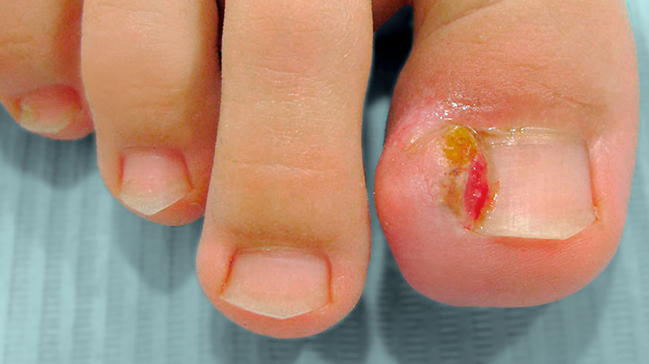
Onychocryptosis (Ingrown toe-nail)
This is a conflict between lamina and periungual tissues, source of pain, morbidity and functional impotence. It is a specific condition of the toenails. The treatments are numerous: quiropodologic and surgical. Surgical management includes two types of therapeutic approach: those aimed at periungual tissues and those concerning the nail tablet, among which, partial avulsion of the tablet followed by phenolization, or Boll's technique is always popular. . |
|
|
|
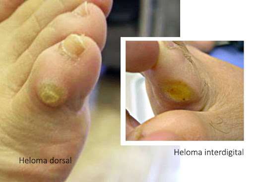
Helomas and hyperkeratosis Helomas (corns) are localized thickenings of the horny layer of the skin, which form a cone-shaped core and often cause pain in the feet. They can be found on the hammer toe joints, between the toes or on the sole of the foot. Hyperkeratosis (calluses) are diffuse thickenings of varying size of the skin, often seen under the forefoot or heel. Both of these foot conditions are often caused by poorly fitting shoes; they can also be due to mechanical instability of the foot which creates friction or repeated overload in the affected area. Dry skin can also be a common initiating factor. |
|
|

|
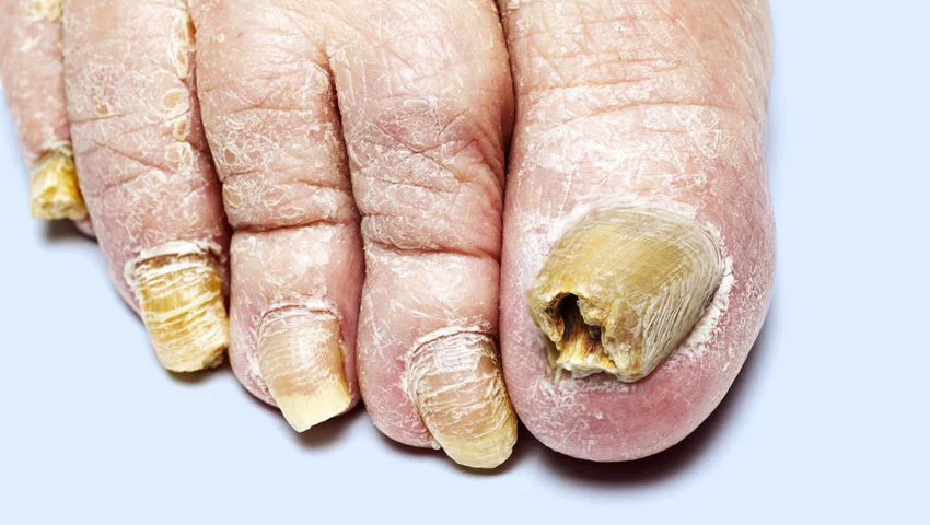
Onychomycosis Onychomycosis, also called nail fungus or nail fungus, is an infection of the nails. Toenail fungus can be caused by several species of microscopic fungi; most often they are dermatophytes (especially on the feet), then yeasts of the Candida genus (especially on the hands) and even more rarely molds (2 to 17% of cases2). The nails most affected are those of the feet, and in particular that of the big toe. Transmission of dermatophytes often appears to be human-to-human. In 2014 in France, onychomycosis accounted for around 30% of superficial mycoses presented to dermatologists, and nearly 50% of the causes of nail pathologies (called “onychopathies”). Removing the dermatophytes responsible for the infection does not restore the normal shape and color of the nail, which only returns with the gradual growth of the nail. In recent years, dermatologists have reported a growth in the number of onychomycosis due to non-dermatophytic molds (NDM), without being able to explain this development. In these cases, the pathogens are, for example, Scopulariopsis brevicaulis or species of the genera Fusarium and Aspergillus or even other rare molds. |
|
|
|
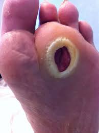
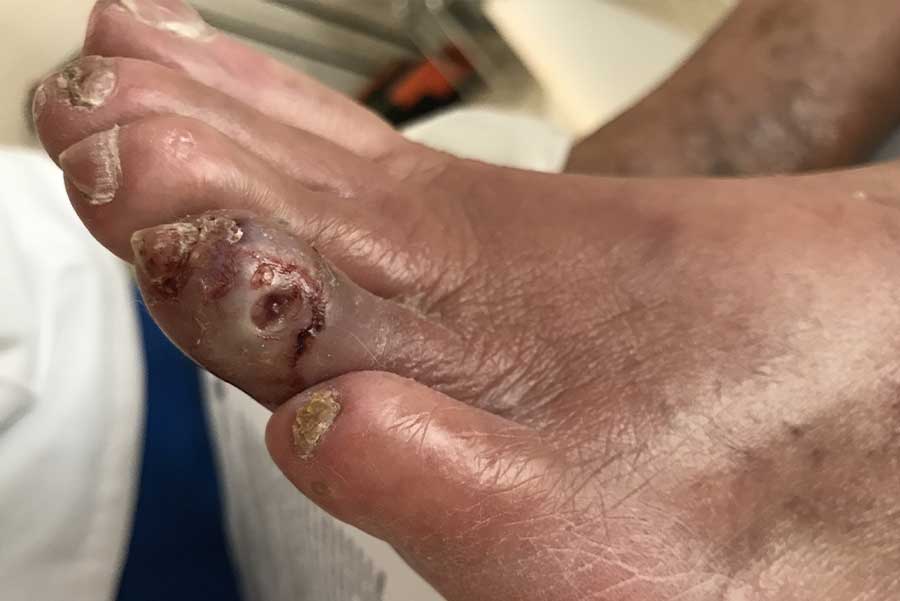
Diabetic foot ulcer: a sneaky ailment In diabetics, the foot ulcer results from 3 mechanisms:
The triggering factors include minor traumas such as unsuitable shoes, injured fingernails, barefoot walking… Due to loss of sensitivity, these superficial injuries are often ignored by the patient. This is why 40% of diabetic foot ulcers are discovered incidentally. Standardized treatment The treatment of diabetic foot ulcer is preferably done in a specialized center. It includes cleaning the wound, making dressings, taking antibiotics and especially a strict discharge. The discharge consists in avoiding the support of the ulcer on the ground. This involves the patient's bed rest, the use of crutches or devices such as orthopedic shoes or plaster. Non – removable landfill casts are the most effective according to Dr Faure, pharmacist at the Montpellier University Hospital. These kinds of resin or plaster boots cover the whole foot and the leg up to the knee. Inside, inflatable pads protect the foot. When walking, the pressures are distributed over the entire arch of the foot and a large part is evacuated upwards due to the rigidity of the system. These boots can be paired with connected socks that detect changes in pressure or temperature. |
|
|
|
Mycosis of the foot or athlete's foot Athlete's foot is by no means a particular aptitude for athletics. This term refers to dermatomycosis, in other words an infection of the skin by a microscopic fungus, which is most often manifested by peeling of the skin and the occurrence of cuts between the toes. Fortunately, effective treatments can overcome it. |
|

|
|
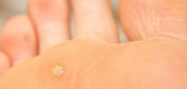
Plantar warts These are benign but contagious tumors, which result from a proliferation of cells in the epidermis. The causative agent is papillomavirus type 1, 2, 4, 7 or 63 (these types are classified on the basis of clinically visible symptoms). |
|
|
|
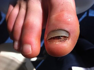
Subungual hematoma An hematoma subungual is an hematoma that is located under a fingernail. It occurs after a trauma or various microtraumas received on the nail |
|
|
|
